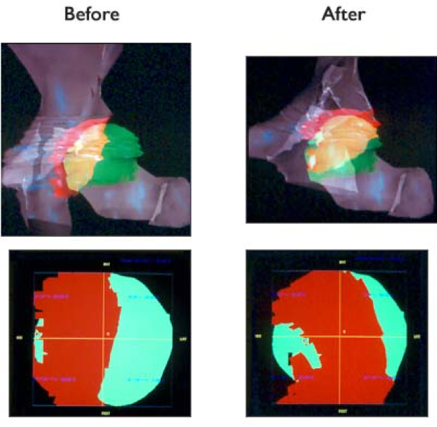
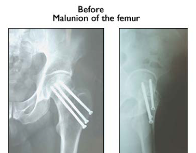
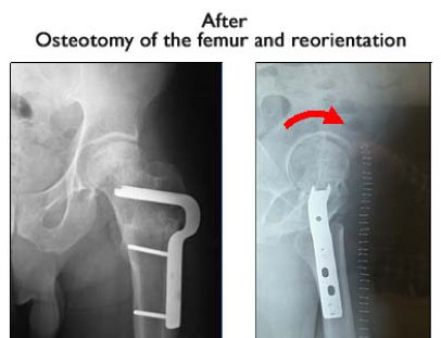
The aim of ReoPlan is to give the orthopaedic surgeon a tool which allows for assessing the relevant morphological factors on three-dimensional imaging. Based on 3D image reconstruction of the hip joint, the tool may predict the feasibility of surgical correction. Osteotomies around the acetabulum and/or about the proximal femur allow for virtual correction of many orthopaedic pathologies.


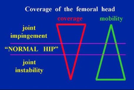
The human hip joint is a ball and socket joint which fulfils two basic tasks by establishing a compromise to achieve normal function: weight bearing and joint mobility. In fact, optimal weight bearing includes a maximal and stable weight bearing surface (coverage) to reduce pressure and shear. Optimal joint mobility includes minimal covering surface (socket) of the head (ball).

Congenital malformation of the hip joint may either include minimal coverage and consequent instability or local overcoverage which causes impingement in selected directions of joint motion. Selected primary or secondary unfavourable shapes of the femoral head may cause impingement too. On the long run, instability and impingement cause joint destruction through osteoarthritis or arthrosis. The coverage of the femoral head is thus a critical parameter to be investigated precisely.


We herewith propose a user friendly programme to be used by the operating surgeon to assess the pathology using conventional and new, more precise quantitative measurements. It furthermore allows for virtual setting of correcting osteotomies of the pelvis and/or the femur and precisely correct the defect by rotating and shifting means.

All correcting means are recorded and can be applied during the surgical intervention. These means include position and lengths of the osteotomies, direction of rotation and shift of the free osteotomized fragment.

Impingement and total range of joint mobility in all directions is recorded too.